Iris in Motion: The Emerging Science of Pupillometry as a Window Into the Brain
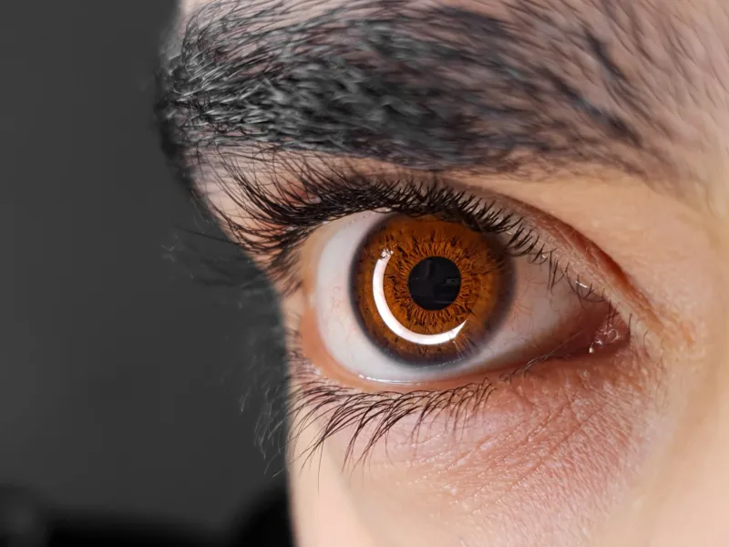
For generations of clinicians, shining a penlight into a patient's eye and observing whether the pupil constricts has served as a rapid, low-cost proxy for neurological integrity. The pupillary light reflex — that involuntary narrowing of the iris in response to illumination — has been a cornerstone of bedside neurological assessment since the earliest days of modern medicine. Yet this familiar reflex represents only a fraction of the information that pupil dynamics can convey. A growing body of research is repositioning pupillometry not as a blunt screening tool but as a sensitive, real-time instrument for probing the functional architecture of the human brain.
Beyond the Light Reflex: A More Complex System
The pupil's size at any given moment is not determined solely by ambient luminance. It is jointly regulated by two branches of the autonomic nervous system — the parasympathetic pathway, which drives constriction via the Edinger-Westphal nucleus, and the sympathetic pathway, which drives dilation. What makes this dual-control system scientifically compelling is that the sympathetic arm is directly modulated by the locus coeruleus (LC), a small nucleus in the brainstem that serves as the brain's primary source of norepinephrine. Because the LC is deeply embedded in arousal, attention, and stress-response networks, changes in its activity are faithfully mirrored in pupil diameter — even in the complete absence of any change in lighting conditions.
This LC-pupil coupling has become a focal point for researchers seeking non-invasive proxies for central nervous system activity. Studies using simultaneous pupillometry and functional MRI have demonstrated robust correlations between LC firing patterns and spontaneous fluctuations in pupil size, lending empirical weight to the idea that the pupil is, in a meaningful sense, broadcasting the brain's internal state to anyone equipped to read it.
Cognitive Load and the Dilating Pupil
Among the most replicated findings in modern pupillometry research is the relationship between mental effort and pupil dilation. When study participants perform tasks of increasing cognitive demand — working memory challenges, mental arithmetic, complex decision-making — their pupils enlarge measurably, even under controlled lighting. This task-evoked pupillary response (TEPR) has been documented across dozens of experimental paradigms and appears to track cognitive load with a granularity that self-report measures cannot match.
Researchers at institutions including Carnegie Mellon University and the University of Amsterdam have used TEPR to distinguish between different types of cognitive processing, map attentional bottlenecks, and even predict individual differences in working memory capacity. In applied contexts, this line of research is informing the design of adaptive interfaces, aviation safety protocols, and human-factors engineering, where real-time assessment of operator mental load could prevent errors before they occur.
Emotional States and Affective Signaling
Pupil dilation is not confined to cognitive engagement; it is equally responsive to emotional arousal. Both positively and negatively valenced stimuli — images that provoke fear, desire, disgust, or joy — trigger measurable pupillary enlargement, reflecting the broader activation of the autonomic nervous system during affective processing. This response is largely independent of stimulus brightness, making it possible to disentangle emotional reactivity from simple luminance effects with appropriately controlled experimental designs.
The implications for clinical psychology are considerable. Researchers have reported atypical pupillary responses to emotionally salient stimuli in individuals with post-traumatic stress disorder, social anxiety disorder, and major depressive disorder. In PTSD, for instance, hyperreactive pupil dilation to threat-related cues has been proposed as a physiological correlate of the hypervigilance that defines the condition. While these findings remain at the translational frontier, they raise the prospect of pupillometry serving as an objective, low-burden complement to subjective symptom inventories in psychiatric assessment.
A Potential Biomarker for Neurodegeneration
Perhaps the most clinically urgent application of advanced pupillometry lies in its potential as an early biomarker for neurodegenerative disease. Several research groups have reported that the pupillary light reflex — specifically its dynamic parameters such as constriction velocity, maximum constriction amplitude, and redilation latency — is measurably altered in patients with Alzheimer's disease, Parkinson's disease, and multiple system atrophy, sometimes preceding the emergence of overt cognitive or motor symptoms.
A 2021 study published in Neurology found that cognitively normal older adults with elevated amyloid burden — a preclinical hallmark of Alzheimer's pathology — displayed subtly impaired pupillary responses compared with amyloid-negative peers, suggesting that pupillometry might one day serve as a low-cost screening adjunct to expensive PET imaging or cerebrospinal fluid analysis. Similarly, research into autonomic dysfunction in Parkinson's disease has identified characteristic pupillometric signatures that reflect the progressive degeneration of noradrenergic and cholinergic circuits.
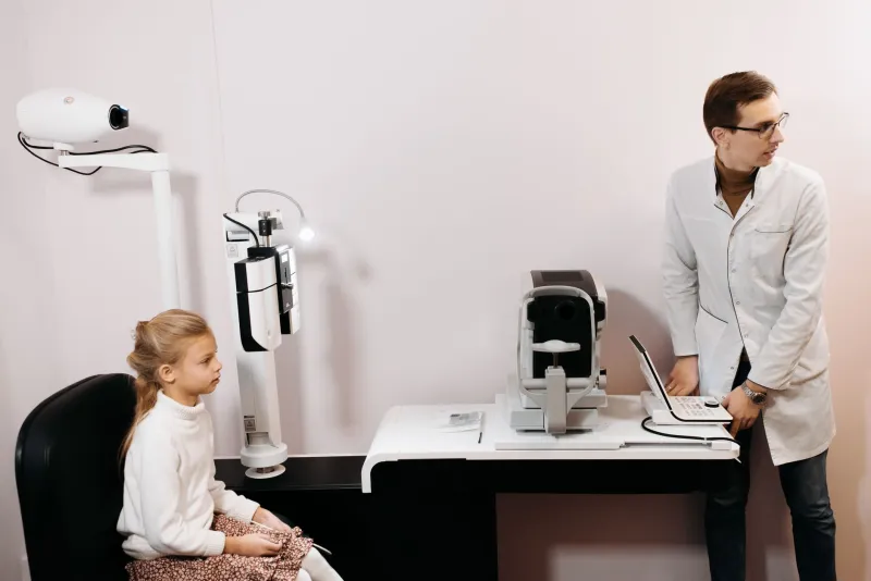
For a healthcare system grappling with the enormous costs of neurodegenerative disease diagnosis, the appeal of a rapid, non-invasive, and inexpensive pupillometric screen is self-evident. The technology required — infrared pupillometers capable of capturing high-resolution iris dynamics — is already commercially available and increasingly portable.
Pupillometry in Psychiatric and Pharmacological Research
Beyond neurodegeneration, pupil dynamics are attracting attention as outcome measures in psychiatric drug trials and basic pharmacology research. Because norepinephrine and acetylcholine — the primary neurotransmitters governing pupil behavior — are also central targets of many psychotropic medications, pupillometry offers a window into the pharmacodynamic effects of drugs at the level of the central nervous system. Dose-response relationships that would otherwise require invasive methods to establish can, in some cases, be approximated through careful pupillometric measurement.
Researchers studying psychedelic-assisted therapy have likewise noted that compounds such as psilocybin and MDMA produce distinctive pupillary signatures during their peak psychoactive phases, opening possibilities for objective monitoring of drug effect in clinical trial settings where subjective self-report is inherently limited.
Methodological Considerations and Limitations
Despite its promise, pupillometry is not without challenges. Pupil size is influenced by a wide array of variables — age, iris pigmentation, refractive state, fatigue, caffeine consumption, and ambient temperature among them — that must be carefully controlled or statistically accounted for in research designs. Individual baseline variability is substantial, and the field has yet to converge on universally accepted normative data sets for clinical reference.
Furthermore, while the correlations between pupil dynamics and brain states are robust at the group level, their sensitivity and specificity at the individual level remain insufficient for standalone diagnostic use in most clinical contexts. Researchers are candid that pupillometry is most powerful as one element within a multimodal assessment framework rather than as an isolated biomarker.
The Diagnostic Horizon
What is becoming clear is that the pupil, long treated as little more than an aperture regulating retinal illumination, is in fact a peripheral readout of some of the brain's most fundamental operating parameters. The convergence of high-resolution infrared imaging, computational analysis of pupil waveform dynamics, and large-scale normative datasets is gradually transforming pupillometry from a laboratory curiosity into a clinically actionable tool.
For vision scientists and neurologists alike, this represents a compelling intersection of ophthalmology and systems neuroscience — one in which the eye's most visible moving part becomes a portal to the invisible machinery of cognition, affect, and neurological health. As the field matures, the routine clinical question of whether a pupil responds to light may give way to a far richer inquiry: precisely how it responds, and what that precision reveals about the brain behind it.