Neural Networks and the Retina: How Machine Learning Is Redefining Disease Detection in Ophthalmology
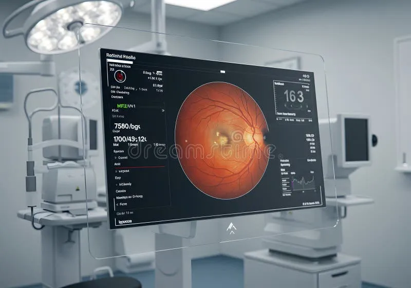
Photo: AI retinal scan analysis ophthalmology laboratory, via thumbs.dreamstime.com
For decades, retinal imaging has served as one of medicine's most revealing windows into systemic and ocular health. The posterior segment of the eye — with its layered architecture of photoreceptors, blood vessels, and neural tissue — encodes a remarkable density of diagnostic information. Historically, extracting that information has required years of subspecialty training. Today, a growing body of research suggests that convolutional neural networks and deep learning frameworks can perform comparable, and in certain conditions superior, pattern recognition on retinal scans — in a fraction of the time.
The implications for vision science are substantial. Researchers, clinicians, and healthcare institutions across the United States are grappling not only with the technical promise of these tools but also with the complex questions they raise about validation standards, workflow integration, and the evolving role of human expertise.
The Algorithmic Turn in Retinal Diagnostics
The foundational shift began gaining serious momentum following a landmark 2016 study published in JAMA by researchers at Google, in collaboration with ophthalmologists at institutions including the University of California, San Francisco. The team trained a deep learning algorithm on over 128,000 retinal fundus images and demonstrated that the system could detect diabetic retinopathy with sensitivity and specificity comparable to board-certified ophthalmologists. That study catalyzed a wave of subsequent research and, eventually, regulatory engagement.
In 2018, the U.S. Food and Drug Administration authorized IDx-DR — the first AI-based diagnostic device cleared for clinical use without requiring a specialist to interpret results. Designed to detect diabetic retinopathy in primary care settings, the system exemplified a new paradigm: AI not as a supplement to specialist review, but as an autonomous first-line screening tool. For underserved communities and rural populations with limited access to retinal specialists, the public health implications are difficult to overstate.
Since then, research into AI-assisted retinal analysis has expanded considerably. Algorithms trained on optical coherence tomography (OCT) images have demonstrated high accuracy in identifying drusen deposits, retinal fluid accumulation, and geographic atrophy — hallmarks of age-related macular degeneration (AMD). A 2020 study from the Moorfields Eye Hospital and DeepMind collaboration, widely referenced in American ophthalmology literature, showed that a deep learning system could recommend appropriate referral decisions for over 50 distinct retinal pathologies with accuracy matching that of expert clinicians.
Methodological Challenges and Validation Standards
Despite these advances, the research community has raised legitimate concerns about the rigor with which AI ophthalmic tools are evaluated before deployment. A systematic review published in The Lancet Digital Health examined 94 studies on AI-based diabetic retinopathy detection and found that the majority were retrospective, relied on limited geographic or demographic diversity in training datasets, and lacked prospective validation in real-world clinical environments.
This matters considerably in a country as demographically diverse as the United States. Retinal pathology can present differently across populations distinguished by age, ethnicity, and comorbidity burden. An algorithm trained predominantly on images from one population cohort may underperform when applied to another — a form of algorithmic bias with direct consequences for patient outcomes. Researchers at institutions including Johns Hopkins and Stanford have begun publishing work specifically examining how dataset composition affects model generalizability, and the National Eye Institute has identified AI fairness as a priority area in its strategic planning documents.
Standardization of performance benchmarks remains another open question. Unlike pharmaceutical trials, which follow well-established protocols under FDA oversight, AI diagnostic tools have entered the market through a regulatory framework — the 510(k) clearance pathway — that some researchers argue is insufficiently rigorous for software that continuously learns and updates. The FDA has acknowledged this gap and published draft guidance on predetermined change control plans for AI/ML-based software, but the scientific community continues to debate what constitutes adequate evidence of clinical validity.
Accelerating Research Workflows Beyond Diagnostics
The impact of machine learning in vision science extends beyond clinical diagnosis. In research settings, AI tools are accelerating the analysis of large-scale imaging datasets in ways that would be prohibitively time-consuming through manual review. Population-level studies examining retinal vascular geometry as a biomarker for cardiovascular disease risk, for instance, have benefited enormously from automated segmentation algorithms that can process thousands of images with consistent precision.
Similarly, AI-assisted analysis of longitudinal OCT datasets is enabling researchers to track subtle structural changes in the retina over time — changes that may precede clinically detectable vision loss by months or years. This capacity for early-stage biomarker identification has particular relevance for conditions like glaucoma and AMD, where intervention timing is closely linked to outcomes. Research groups at the University of Pennsylvania and the Wilmer Eye Institute at Johns Hopkins are among those leveraging these capabilities in ongoing longitudinal studies.
Generative AI models are also beginning to appear in vision research contexts, used to augment training datasets with synthetic retinal images — a potential solution to the persistent problem of limited annotated data for rare conditions. While promising, this approach introduces its own validation challenges, as the fidelity and clinical representativeness of synthetic imagery must be rigorously established before such data can responsibly inform diagnostic algorithms.
The Question of Human Expertise in an AI-Augmented Field
Perhaps the most philosophically significant question emerging from this body of research concerns the future relationship between machine intelligence and human clinical expertise. Some commentators have predicted that AI will eventually supplant retinal specialists in routine screening contexts. Most researchers and clinicians, however, articulate a more nuanced view: that AI will function most effectively as a collaborative instrument — amplifying the diagnostic capacity of trained professionals rather than replacing their judgment.
There is evidence to support this framing. Studies examining human-AI collaborative diagnostic performance have generally found that the combination of algorithmic screening and specialist review outperforms either approach in isolation, particularly for ambiguous or complex cases. The subspecialty of retinal imaging may be entering an era defined not by competition between human and machine cognition, but by the careful orchestration of both.
For researchers in vision science, this moment demands both intellectual openness and methodological discipline. The tools now available are genuinely powerful. Their responsible integration into clinical and research practice will depend on the rigor with which the scientific community examines their limitations, advocates for equitable validation standards, and maintains a commitment to patient-centered outcomes as the primary measure of success.
