Beyond the Visible: How Research Into UV and Infrared Perception Is Opening New Frontiers in Vision Science

Photo by Photo by Marek Pavlík on Unsplash on Unsplash
The human eye is, by any reasonable measure, a remarkable instrument. Yet it perceives only a sliver of the electromagnetic energy that surrounds it — wavelengths spanning roughly 380 to 700 nanometers, a range we call visible light. Below that threshold lies the ultraviolet spectrum; above it, infrared radiation. For most of human history, these adjacent regions were simply invisible — not absent, but inaccessible. What is now becoming clear, through an accelerating body of interdisciplinary research, is that the boundary between visible and invisible is neither as fixed nor as irrelevant to human health as once believed.
Researchers in vision science, photobiology, and biomedical optics are finding that ultraviolet (UV) and infrared (IR) wavelengths interact with the visual system — and with biological tissue more broadly — in ways that carry significant clinical and diagnostic implications. The work is reshaping how scientists think about the evolutionary architecture of human sight, and it is pointing toward practical applications that range from enhanced ophthalmic imaging to entirely new treatment modalities for retinal disease.
Rethinking the Visible Spectrum's Boundaries
The conventional account of human spectral sensitivity rests on the absorption properties of three classes of cone photoreceptors, each tuned to peak sensitivity in the short (blue), medium (green), and long (red) wavelength ranges. Ultraviolet light, which falls below 380 nanometers, is largely filtered by the cornea and crystalline lens before it can reach the retina — a protective mechanism that prevents phototoxic damage to photoreceptor cells. This filtering has long been interpreted as evidence that UV sensitivity is simply not part of the human visual repertoire.
Recent research has complicated that narrative in instructive ways. Studies examining patients who have undergone cataract surgery and received certain intraocular lens (IOL) implants — particularly older UV-transmissive designs — have reported anecdotal sensitivity to near-UV wavelengths. More rigorously, psychophysical experiments have documented that the short-wave cone system retains residual sensitivity into the near-UV range when the natural lens is absent or replaced. A notable case study frequently cited in this literature involved a visual artist who, following aphakia, reported perceiving ultraviolet light as a pale blue-white hue — a perceptual experience consistent with S-cone stimulation at sub-visible wavelengths.
These findings do not suggest that typical human vision is UV-sensitive under ordinary conditions. Rather, they reveal that the biological machinery for near-UV detection may be latent within the photoreceptor system, suppressed primarily by the optical filtering properties of the anterior eye. For researchers, this distinction is significant: it suggests that the spectral limits of human vision are partly architectural rather than purely neurobiological.
Infrared Research and the Emerging Science of Photobiomodulation
On the opposite end of the near-visible spectrum, infrared light — particularly the near-infrared (NIR) range between 700 and 1,400 nanometers — has become a subject of intense research interest, driven in part by the emerging field of photobiomodulation (PBM). PBM refers to the therapeutic application of low-level light at specific wavelengths to modulate cellular function, and it has attracted considerable attention as a potential intervention for retinal degenerative diseases.
The mechanistic basis for PBM in retinal tissue centers on the absorption of near-infrared light by cytochrome c oxidase, a mitochondrial enzyme central to cellular energy production. Research conducted at institutions including University College London and, in the United States, at the Bascom Palmer Eye Institute, has examined whether brief, low-intensity NIR exposure can enhance mitochondrial function in aging photoreceptors — cells that are among the most metabolically demanding in the human body.
A randomized controlled trial published in Journals of Gerontology in 2021, led by a team at University College London, found that three minutes of daily 670-nanometer light exposure improved color contrast sensitivity in participants over 40, with effects persisting for at least one week following treatment. While the study was modest in scale, it generated significant discussion within the American vision research community and has prompted follow-on investigations exploring dose-response relationships and longer-term outcomes in older populations.
For researchers working on age-related macular degeneration and other conditions characterized by mitochondrial dysfunction in retinal pigment epithelium cells, these findings represent a potentially important therapeutic avenue — one that, notably, operates through a spectral channel entirely outside the range of ordinary human visual experience.
UV and IR as Diagnostic Instruments
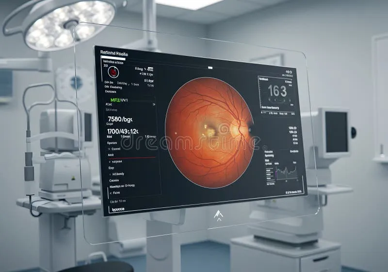
Beyond their therapeutic potential, UV and infrared wavelengths have long been employed as diagnostic tools in ophthalmic imaging — though the sophistication of these applications has grown considerably in recent years. Fundus autofluorescence (FAF) imaging, which exploits the natural fluorescent properties of lipofuscin in the retinal pigment epithelium when excited by blue or near-UV light, has become a standard modality for monitoring the progression of conditions including AMD, Stargardt disease, and various retinal dystrophies.
Near-infrared reflectance imaging, meanwhile, offers complementary structural information about the deeper layers of the retina and choroid, with reduced scattering artifacts compared to shorter wavelengths. Multimodal imaging platforms that integrate visible, near-UV, and near-IR channels are increasingly standard in academic ophthalmology centers across the United States, enabling researchers to construct more comprehensive maps of retinal pathology than any single imaging modality permits.
Perhaps the most technically ambitious development in this space is the application of infrared adaptive optics systems to retinal imaging. By correcting for the wave-front aberrations introduced by the eye's optical components, adaptive optics allows researchers to image individual photoreceptor cells in living subjects — a resolution that was unattainable just two decades ago. When combined with infrared illumination sources, these systems minimize phototoxic risk while preserving imaging fidelity, making longitudinal cellular-level studies more feasible in human research participants.
Evolutionary Constraints and Their Clinical Relevance
The question of why human vision evolved to occupy its particular spectral niche is not merely academic. Understanding the selective pressures that shaped our photoreceptor complement — and the trade-offs those pressures imposed — has direct implications for interpreting vulnerability to phototoxic damage, designing protective interventions, and evaluating the biological plausibility of proposed spectral therapies.
Comparative research across vertebrate species offers instructive context. Many birds, fish, and insects possess UV-sensitive photoreceptors as a functional component of their visual systems, enabling behaviors — mate selection, foraging, navigation — that depend on UV pattern discrimination. Humans and other catarrhine primates lost UV sensitivity at the receptor level during evolutionary history, a shift linked to trichromatic color vision and the filtering adaptations that protect the macula from short-wavelength phototoxicity.
This evolutionary framing helps clarify why the intersection of UV and IR research with human health is so consequential. The same spectral regions that our visual systems are designed to exclude are, paradoxically, among the most biologically active in terms of their interactions with ocular tissue. Ultraviolet exposure is a well-established risk factor for cataract formation, pterygium, and photokeratitis. Near-infrared radiation, while less acutely damaging, has been implicated in thermal injury to the lens and retina at high intensities. Understanding these interactions at a mechanistic level is essential for developing rational photoprotection strategies and for calibrating the therapeutic applications of these wavelengths safely.
The research field positioned at the intersection of spectral biology, ophthalmic imaging, and photomedicine is still maturing. But the convergence of advanced optical instrumentation, improved mechanistic understanding, and renewed clinical interest in non-pharmacological retinal therapies suggests that UV and infrared vision science will occupy an increasingly prominent place in the broader landscape of vision research — and in the clinical toolkit available to practitioners working to preserve and restore human sight.