The Gap That Isn't There: What the Brain's Physiological Blind Spot Reveals About the Architecture of Perception
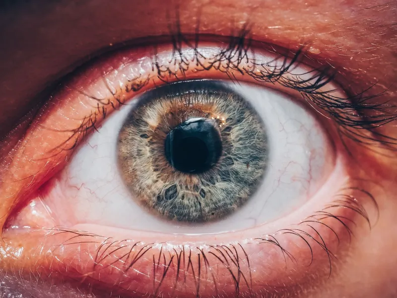
Somewhere in your right visual field, at this very moment, there is a region roughly the size of a full moon that your right eye cannot see. The same is true of your left eye in the opposite hemifield. These are not metaphorical blind spots—they are anatomical facts, the direct consequence of the optic nerve's exit point from the retina, a location known as the optic disc. At this junction, no photoreceptors exist. No rods. No cones. Nothing capable of transducing light into neural signals.
And yet, for the overwhelming majority of people, no corresponding void appears in conscious experience. The world looks continuous, seamless, and complete. This is not a trivial observation. It is, according to a growing body of neuroscientific research, one of the most instructive demonstrations available to vision scientists—a built-in experiment that the nervous system runs on itself, continuously, without pause.
Anatomy of an Absence
The optic disc, situated approximately 15 degrees temporal to the fovea in each eye, represents the point at which roughly one million retinal ganglion cell axons converge and exit the eye to form the optic nerve. Because this region is occupied entirely by axonal fibers rather than photoreceptors, it creates a scotoma—a localized area of absent visual function—in each monocular visual field.
The dimensions of this scotoma are not trivial. Measured in degrees of visual angle, the physiological blind spot spans approximately 5 to 7 degrees vertically and 7 to 9 degrees horizontally. At arm's length, this corresponds to a region large enough to obscure a human face. The fact that this absence rarely intrudes into conscious awareness is, from a neuroscientific standpoint, the central mystery worth examining.
It is important to distinguish this from the colloquial use of "blind spot" in psychological discourse. The physiological blind spot is a hard anatomical constraint, not a cognitive bias or attentional lapse. Its study therefore offers researchers a relatively clean experimental handle on a question that is otherwise difficult to isolate: how does the visual brain manage missing information?
Filling In: More Than Meets the Eye
The process by which the brain compensates for the blind spot is commonly described in the literature as "filling in" or perceptual completion. But this language, while convenient, risks understating the sophistication of the underlying mechanisms.
Early theoretical accounts proposed that filling in was essentially passive—a simple interpolation based on surrounding visual information, analogous to a digital photo editor's content-aware fill tool. More recent experimental work has complicated this picture considerably. Neuroimaging studies, including those using high-resolution functional MRI, have demonstrated that the cortical region corresponding to the blind spot—a specific portion of primary visual cortex (V1)—does not simply go dark in the absence of retinal input. Instead, it receives active signals, apparently driven by top-down feedback from higher visual areas.
This finding carries significant theoretical weight. It suggests that filling in is not merely an absence of signal but a positive neural event—the brain is not simply ignoring the gap; it is generating content to occupy it. Whether this constitutes genuine perceptual experience or a pre-conscious process that never reaches awareness remains an active area of debate among researchers working in the philosophy and neuroscience of consciousness.
What Surrounds the Void Determines What Fills It
One of the more experimentally productive aspects of blind spot research is the degree to which filling-in content can be manipulated by controlling the visual surround. Classic psychophysical experiments have demonstrated that when a uniform texture or color pattern surrounds the blind spot region, subjects perceive that pattern as continuous across the scotoma. When a moving stimulus surrounds the blind spot, motion perception can propagate into the region. These findings have been replicated and extended across numerous laboratory settings.
More recent work has examined the temporal dynamics of filling in, asking how quickly the brain completes the gap following changes in the surrounding image. The answer—measured in tens to hundreds of milliseconds depending on stimulus complexity—reveals that completion is not instantaneous and that the underlying computation is non-trivial. Researchers have also demonstrated that filling in can fail under certain conditions, particularly when high-contrast or rapidly changing stimuli are presented at the blind spot boundary, allowing subjects to briefly perceive a dark or ambiguous region in their visual field.
This conditionality is scientifically valuable. It allows investigators to probe the limits of the completion mechanism and to identify the variables that determine its success or failure, thereby mapping the computational constraints of the underlying neural circuitry.
A Window Into Predictive Processing
The blind spot has become a touchstone for researchers working within the predictive processing framework of perception—a theoretical paradigm that has gained substantial traction in cognitive neuroscience over the past two decades. In this framework, the brain is understood not as a passive receiver of sensory information but as an active prediction machine that continuously generates expectations about incoming signals and updates those predictions based on discrepancies, or prediction errors.
From this perspective, filling in the blind spot is not an anomaly requiring special explanation. It is simply what the visual brain does all the time—generate a best-fit model of the world based on available evidence, prior experience, and statistical regularities in the environment. The blind spot merely makes this process visible to scientific inquiry because the absence of sensory input is so absolute and anatomically defined.
This interpretive lens has implications well beyond the blind spot itself. If the brain's default mode is predictive construction rather than faithful image capture, then the seamless visual world we experience is, in a rigorous sense, a controlled hallucination—a continuously updated model that happens, under normal circumstances, to correspond reliably to external reality. The blind spot is one of the rare instances where the seam in this construction can be experimentally exposed.
Clinical and Research Implications
Beyond its theoretical significance, blind spot research has practical applications in clinical vision science. Accurate mapping of the physiological blind spot and its boundaries is a component of visual field testing, and changes in the apparent size or position of the blind spot can serve as indicators of pathological processes affecting the optic nerve or peripapillary retina, including early-stage glaucoma.
Researchers have also explored whether individual differences in the efficiency of blind spot filling in correlate with broader visual processing traits or with susceptibility to certain visual illusions. Preliminary findings suggest such correlations may exist, though the literature remains insufficiently large to draw firm conclusions. This represents a productive avenue for future investigation, particularly as neuroimaging techniques continue to improve in spatial and temporal resolution.
Additionally, computational models developed to explain blind spot completion have informed the design of artificial vision systems, including image inpainting algorithms used in machine vision and digital media applications. The cross-pollination between biological and artificial vision research in this domain exemplifies the broader scientific value of studying perceptual phenomena at the boundary of sensory capacity.
Rethinking What Vision Is
The physiological blind spot occupies a peculiar position in the landscape of vision science. It is simultaneously one of the most well-known and least-examined phenomena in the field—familiar enough to appear in introductory textbooks, yet rich enough to support sophisticated experimental and theoretical work at the frontier of perceptual neuroscience.
What it ultimately reveals is that vision, as experienced, is not a direct readout of the retinal image. It is a construction—a neural synthesis that integrates sensory evidence with prior knowledge, contextual inference, and active prediction. The gap in the retina that the brain so reliably and invisibly fills is, in this sense, not a flaw in the visual system. It is a feature: one that, properly examined, discloses the architecture of perception with unusual clarity.