Damage Before Darkness: The Case for Structural-First Glaucoma Detection and Why Clinics Haven't Caught Up
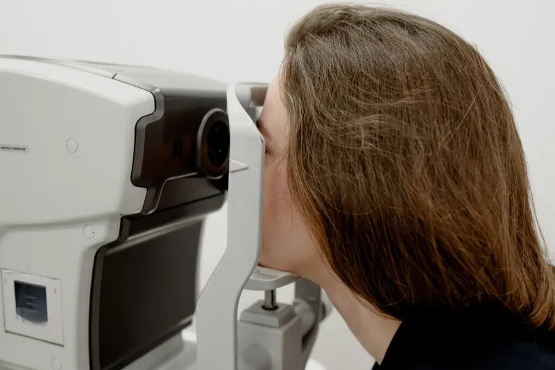
Glaucoma is frequently described as the silent thief of sight, a phrase that captures its most clinically troubling characteristic: the disease causes irreversible damage to the optic nerve across a timeline that renders patient-reported symptoms nearly useless as early indicators. By the time a patient presents with noticeable visual field loss — the functional deficit that traditionally triggers or confirms a glaucoma diagnosis — retinal ganglion cell loss has typically reached 20 to 40 percent. That is not a detection system. That is a documentation system for damage already done.
The scientific tools to do better exist. The clinical infrastructure to deploy them broadly does not — at least not yet. Examining that gap, and the structural and economic forces that sustain it, is as important to the future of glaucoma care as any laboratory breakthrough.
The Structural-Functional Divide
At the heart of glaucoma's diagnostic challenge lies a well-documented dissociation between structural and functional measures of disease severity. Standard automated perimetry — the visual field test that remains the functional cornerstone of glaucoma monitoring in the United States — detects loss only after it has become statistically significant relative to age-matched normative data. The threshold for detection is high, and the variability of the test is substantial, often requiring multiple examinations before a change is deemed clinically reliable.
Structural measures, by contrast, can capture tissue-level changes at far earlier stages. Optical coherence tomography (OCT) of the retinal nerve fiber layer and ganglion cell complex has become standard in glaucoma practices over the past two decades, and it consistently detects thinning that precedes functional field loss in longitudinal study populations. The structural deficit comes first; the functional deficit follows. This sequence is not controversial in the research literature. Its implications for diagnostic practice, however, remain incompletely absorbed.
The reason is partly technical. OCT measurements have a floor effect — once retinal nerve fiber layer thickness falls below the instrument's resolution threshold, structural measures lose sensitivity precisely when functional measures are becoming most informative. This creates a crossover point in disease progression that has sometimes been misread as evidence that structural and functional measures are interchangeable across the disease spectrum. They are not. They are complementary, with structural tools offering the greatest advantage in the early stages that matter most for preserving vision.
What Advanced Imaging Reveals at the Nerve Head
The optic nerve head — the site where retinal ganglion cell axons exit the eye en route to the lateral geniculate nucleus — is a mechanically complex structure whose behavior under elevated intraocular pressure is far more nuanced than a simple compression model would suggest. Lamina cribrosa morphology, peripapillary scleral stiffness, and the biomechanical properties of the neural canal all modulate how pressure-related stress is transmitted to axons, and individual variation in these parameters is substantial.
Advanced OCT platforms with enhanced depth imaging can now resolve lamina cribrosa structure with sufficient fidelity to detect changes in curvature, thickness, and focal defects that precede measurable nerve fiber layer loss. Prospective studies have documented lamina cribrosa deformation in ocular hypertensive eyes — those with elevated pressure but no functional deficit — years before those eyes meet conventional criteria for glaucoma conversion. These structural changes are not artifacts. They represent the earliest detectable signature of mechanical vulnerability in the optic nerve head.
Beyond OCT, swept-source imaging modalities offer improved penetration through the sclera and choroid, enabling three-dimensional reconstruction of the posterior pole that reveals tissue-level changes invisible to earlier-generation instruments. Adaptive optics imaging, still largely confined to research settings, can resolve individual retinal ganglion cells and their axonal projections, providing a cellular-resolution view of early degeneration that standard clinical imaging cannot approach.
Electrophysiology: Functional Signatures Before Subjective Loss
If structural imaging captures the anatomical substrate of early glaucomatous injury, electrophysiological testing can detect its functional correlate — altered neural signaling — before it rises to the level of perceptible vision loss. Pattern electroretinography measures the electrical response of retinal ganglion cells to structured visual stimulation and has demonstrated sensitivity to ganglion cell dysfunction in preperimetric glaucoma populations with reasonable specificity.
Frequency-doubling technology perimetry, which targets the magnocellular pathway particularly vulnerable to early glaucomatous damage, similarly detects functional deficits that fall below the threshold of standard automated perimetry. Multifocal electroretinography variants capable of spatially mapping ganglion cell function across the visual field add a topographic dimension to electrophysiological assessment that is directly relevant to clinical decision-making.
None of these tools is perfect. Each has characteristic limitations in sensitivity, specificity, and inter-session variability. But when integrated with structural imaging in a multimodal diagnostic framework, they offer a substantially more complete picture of early glaucomatous disease than any single modality alone — and a picture that arrives far earlier in the disease course than functional perimetry.
The Adoption Gap and Its Causes
Given the available evidence, why do most American glaucoma patients still receive their primary diagnostic workup through tools designed to detect damage after it has become functionally significant? The answer involves a convergence of reimbursement structures, practice economics, regulatory inertia, and the inherent conservatism of clinical medicine in the face of incomplete long-term outcome data.
Medicare reimbursement schedules — which govern payment for a large proportion of the glaucoma-age population in the United States — have been slow to incorporate newer imaging modalities at rates that reflect their diagnostic value. Adaptive optics imaging, for instance, has no established reimbursement pathway, effectively restricting its use to research settings regardless of its clinical potential. Even for modalities with existing billing codes, reimbursement rates have not kept pace with instrument acquisition and maintenance costs, creating economic disincentives for broad adoption in community ophthalmology practices.
There is also a legitimate scientific concern: demonstrating that earlier structural detection translates into meaningfully better long-term visual outcomes requires large, long-duration randomized trials that have not yet been completed. Regulatory and payer bodies reasonably ask for outcome evidence before mandating or reimbursing diagnostic change. That evidence is accumulating but remains incomplete, sustaining a holding pattern that serves neither patients nor the field.
Toward a Structural-First Diagnostic Paradigm
The scientific case for restructuring glaucoma diagnosis around structural vulnerability rather than functional confirmation is compelling. The optic nerve does not announce its distress through symptoms. It communicates through tissue-level changes that imaging can now read with increasing precision. A diagnostic system calibrated to hear that communication — rather than waiting for the patient to report what they can no longer see — is not merely technologically feasible. It is, by the available evidence, the scientifically defensible standard toward which the field should be moving.
Achieving that transition will require coordinated effort across research, regulatory, and clinical domains: prospective outcome trials that link early structural detection to preserved visual function, reimbursement reform that removes economic barriers to advanced imaging adoption, and training initiatives that prepare clinicians to interpret and act on structural data before functional thresholds are crossed. The science has outpaced the system. Closing that gap is the field's most pressing translational challenge.